Infective Endocarditis: Difference between revisions
No edit summary |
No edit summary |
||
| Line 174: | Line 174: | ||
#iii Miró, José M, Ana del Rı́o, and Carlos A Mestres. "Infective endocarditis in intravenous drug abusers and HIV-1 infected patients." Infectious disease clinics of North America 16.2 (2002): 273-295. | #iii Miró, José M, Ana del Rı́o, and Carlos A Mestres. "Infective endocarditis in intravenous drug abusers and HIV-1 infected patients." Infectious disease clinics of North America 16.2 (2002): 273-295. | ||
#iv Ipek, Esra Gucuk et al. "Infections of implantable cardiac rhythm devices: predisposing factors and outcome." Acta cardiologica 67.3 (2012): 303. | #iv Ipek, Esra Gucuk et al. "Infections of implantable cardiac rhythm devices: predisposing factors and outcome." Acta cardiologica 67.3 (2012): 303. | ||
#v | #v pmid=23720451 | ||
#vi pmid=21685200 | #vi pmid=21685200 | ||
#vii | #vii pmid=22624837 | ||
#viii pmid=16028160 | #viii pmid=16028160 | ||
#x pmid=16767483 | #x pmid=16767483 | ||
#xi pmid=19665088 | #xi pmid=19665088 | ||
Revision as of 18:17, 3 December 2013
Introduction
Infective endocarditis (IE) is an infectious and inflammatory process of endothelial lining of the heart structures and valves. It is most commonly caused by bacterial and fungal infections, although non-infective causes of endocarditis occur, this chapter will concentrate on infective causes.
Epidemiology
The global incidence of endocarditis varies in literature with a wide range of 3 to 9 cases per 100,000 person years[1]. Although it appears that in the elderly population the incidence rate is remarkably higher reaching 20.4 cases per 100,000 person years[2]. This is likely related to increased hospitalization, valve replacement surgeries and intra-cardiac instrumentation in the elderly population. In addition to aging, the prevalence of chronic diseases predisposing to bacteremias such as human immunodeficiency syndrome, end-stage renal disease, organ transplantations and diabetes have also increased. IE is a dreaded complication of intravenous drug use, most commonly affecting the tricuspid valve, with a yearly incidence of 1-5% among chronic users[3]. IE related to implantable rhythm devices remains relatively low but is on the rise due to increased patient population requiring rhythm devices[4].
Pathogenesis and Causes
Generally, endothelial damage to heart valves predisposes to bacterial infections as it is generally resistant to bacterial infections. This may be caused by turbulent blood flow in damaged valves, septal defects or instrumentation. Although, recent evidence suggests that A-type von Willebrand factor may contribute to S. aureus binding in endothelial intact valves[5]. Specifically, in patients with S. aureus bacteremia, native valve endocarditis was reported to be in 19% of patients, and 38% in those with prosthetic valves[6].
While many microorganisms have been implicated in endocarditis syndromes, few infectious bacteria account for the majority of cases.
Staphylococcal endocarditis is most commonly caused by S.aureus. It is the most lethal organism implicated in endocarditis with mortality rates approaching 37%[7]. S.aureus is a highly virulent organism and may cause significant valve destruction, abscess formation, conduction abnormalities and embolization. It often enters the bloodstream from the nares or skin. Patients with left sided involvement often require surgery. In intravenous drug users is the most common cause of IE[8]. Patients who are found to have Staphylococcal bacteremia should undergo echocardiography to rule out endocarditis. The prevalence of endocarditis in patients with S.aureus bacteremia was reported in 19% and 38% in those with native and prosthetic valves respectively[9].
S. epidermidis is an important cause of prosthetic valve endocarditis and is associated with a particularly high incidence of heart failure and valvular abscess formation and a mortality rate of 36%[10].
Viridans group streptococcus often account for 30% of community acquired native valve endocarditis[11]. They are part of the oral cavity flora and may gain entry into the bloodstream via dental caries or trauma. The virulence is generally low and eradication rates are high.
Streptococcus bovis is part of the lower gastrointestinal and urinary tract and is commonly implicated in underlying colorectal disease if found to be the cause of endocarditis. Patients who are found to have S.bovis endocarditis should undergo a colonoscopy to exclude colorectal malignancy or polyps.
Enterococcal endocarditis is part of the gastrointestinal and genitourinary flora and is often implicated in patients in patients undergoing genitourinary or gastrointestinal procedures. Enterococcal endocarditis is generally difficult to treat due to high rates of antibiotic resistance and often require multi-drug regimen.
Gram negative bacilli IE is rather uncommon, the HACEK organisms (Haemophillus spp, Actinobacillus, Cardiobacterium hominis, Eikenella corrodens, Kingella spp) are responsible for approximately 3% of endocarditis cases and are the most common cause for gram negative endocarditis in non-intravenous drug users[12]. Non-HACEK organisms are a rare cause for endocarditis and only account for <1-2% of causes.
Fungal endocarditis occurs in patients who receive prolonged parenteral nutrition or antibiotics through intravenous catheters. It has also been described in intravenous drug users. Patients are often immunocompromised. The most common organisms implicated are Candida species, Histoplasma capsulatum, and Aspergillus. Mortality rates associated with fungal endocarditis exceed 80%[13].

|
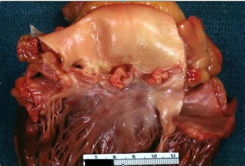
| Large lesions on non coronary and left coronary cusps normal valves otherwise |
|---|

|
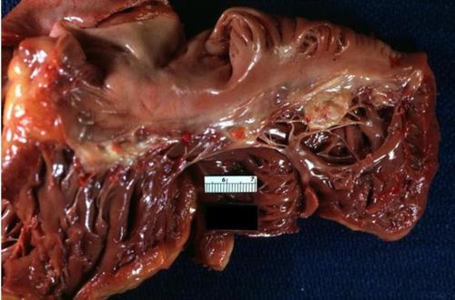
| Vegetations on tricuspid valve |

|
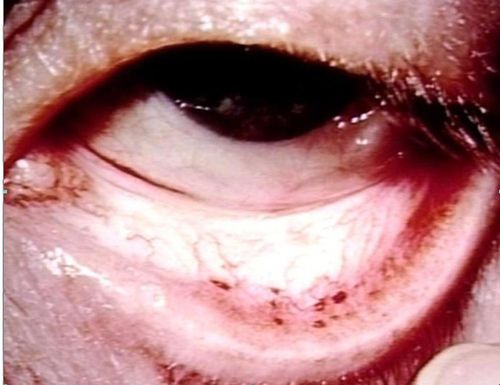
| Septic emboli to the conjunctiva |
Diagnosis
Several diagnostic criteria have been proposed for the diagnosis of IE. In clinical practice, it is the global clinical picture that leads to decision making in the diagnosis and treatment of endocarditis. The modified DUKE criteria for diagnosis is often widely used, with a sensitivity and specificity approaching ~80%.[14] The DUKE criteria divides into Definite IE, Possible IE, or Rejected IE. It uses Major criteria (microbiology, valvular abnormalities) and Minor criteria (systemic symptoms described below). Using the diagnostic criteria for IE should not override clinical judgment.
Definite IE:
- Pathologic Criteria:
- Microorganisms demonstrated by culture or histologic examination of vegetation, a vegetation that has embolized, or an intracardiac abscess specimen OR pathologic lesions; vegetation or intracardiac abscess confirmed by histologic examination showing active endocarditis
- Clinical Criteria
- 2 major criteria OR 1 major and 3 minor criteria OR 5 minor criteria
Possible IE:
- 1 Major criterion + 1 Minor criterion OR 3 minor criteria
Rejected IE:
- Firm alternate diagnosis explaining evidence of IE OR
- Resolution of IE syndrome with =<4 days of antibiotics therapy OR
- No pathologic evidence of IE at surgery or autopsy with antibiotic therapy for =<4 days OR
- Does not meet criteria for IE, as above
| Major Criteria (microbiology) | |
|---|---|
| Typical organisms x 2 blood cultures | e.g S. viridans, S. bovis, HACEK (Haemophilus spp, Aggregatibacter, Cardiobacterium hominis, Eikenella spp, Kingella kingae), S. aureus, Enterecoccus with no primary source |
| Persistent bacteremia | With blood cultures drawn >12 hours apart OR 3 out of 3, or 3 out of 4 positive blood cultures |
| Single positive blood culture for Coxiella burnetti or antiphase I IgG antibody titer >1:800 | |
| Major Criteria (valve) | |
| Echocardiogram with evidence of vegetation | TTE or TEE |
| New valvular regurgitation | |
| Minor Criteria | |
| Predisposing cardiac condition or IV drug use | |
| Fever | >38 degrees celsius or
>100.4 fahrenheit) |
| Vascular phenomena | major arterial emboli, septic pulmonary infarcts, mycotic aneurysm, intracranial hemorrhage, conjunctival hemorrhage, janeway lesions |
| Immune phenomena | glomerulonephritis, Osler nodes, Roth spots, positive RF |
| Positive blood culture not meeting above major criteria or serological evidence of active infection with organism consistent with IE | |
The sensitivity of detecting on echocardiogram varies. Transthoracic and transesophageal echocardiogram sensitivities for detecting vegetations are 50% and 90% respectively.[15]
| Endocarditis of prosthetic mitral valve | |
|---|---|
| http://www.echopedia.org/images/2/24/E00404.swf | http://www.echopedia.org/images/6/6a/E00405.swf |
| Endocarditis of the aortic valve | |
| http://www.echopedia.org/images/1/10/E00114.swf | http://www.echopedia.org/images/5/5b/E00117.swf |
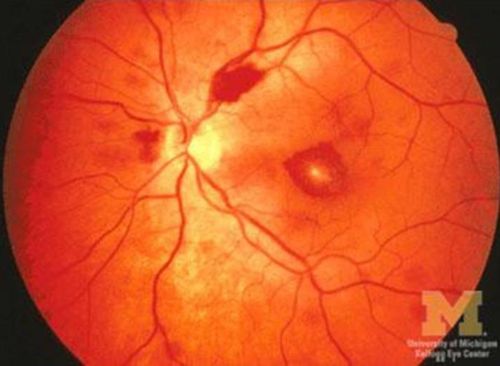
| Roth Spots |
|---|

|
Therapy
Empiric therapy for infective endocarditis should only be used when there is a high index of suspicion in the absence of positive blood cultures. Three blood cultures should be drawn 30 minutes apart prior to initiating treatment. When there a high clinical probability of infective endocarditis in acute settings, empiric therapy is geared towards MRSA and coagulase negative staphylococcus. In healthcare settings and in injection drug users coverage should also include gram negative bacilli. Options for such therapy include Vancomycin + Gentamicin or, Nafcillin/Oxacillin + Tobramycin/Gentamicin.
When considering coverage for subacute endocarditis, coveraged is geared more towards streprococci spp. Options commonly include Ampicillin/Sulbactam + Gentamicin/Tobramycin, or Ceftriaxone + Vancomycin.
As soon as blood cultures become available antibiotics should be adjusted to target the identified microorganisms.
In cases of prosthetic valve endocarditis (PVE), microbiological activity depends on early (<2 months post op) or late (>2 months post op). In early PVE S.aureus accounts for 40% of the cases, followed by coagulase negative staphylococcus (17%). In late PVE coagulase negative staphylococcus accounts for 20% of cases, followed by S. aureus (18%). Coverage for enterococci, streptococci, and gram negative should be considered in empiric therapy in both groups. Rifampin + Vancomycin + Gentamicin should be initiated for PVE <12 post op. Suspected PVE >12 months post op may be treated with the same regimen as for native valves.[16]
The American Heart Association recommendation for specific antimicrobial therapy is shown below: http://circ.ahajournals.org/content/111/23/3167.full
The guidelines on treatment of endocarditis from the task force for treatment of Infective endocarditis of the European Society of Cardiology is shown below: http://eurheartj.oxfordjournals.org/content/30/19/2369.long
Prophylaxis
According the American Heart Association guidelines published in 2007 the following groups of patients are considered to be high-risk and require prophylaxis:[17]
- Any prosthetic heart valve, or prosthetic material used for valve repair
- Previous infective endocarditis
- Congenital heart disease (CHD)
- Unrepaired cyanotic CHD, including all palliative shunts and conduits
- Completely repaired congenital heart defect with prosthetic material or device, whether placed by surgery or by catheter intervention, during the first 6 months after the procedure
- Repaired CHD with residual defects at the site or adjacent to the site of a prosthetic patch or prosthetic device (which inhibit endothelialization)
- Cardiac transplantation recipients who develop cardiac valvulopathy
- Dental procedures require prophylaxis:
- Manipulation of gingival tissue, or the periapical region of teeth or perforation of oral mucosa
- Respiratory tract procedures require prophylaxis:
- Incision or biopsy of the respiratory mucosa, or procedures involving treatment of abscess of empyema
Antibiotic prophylaxis is also recommended for any procedures on infected skin/skin structures or musculoskeletal tissue in high risk patients.[17]
Complications of Endocarditis
Common complications arising from IE can be divided into local and systemic. Local complications often arise from direct extension of the infection into cardiac structures. Systemic complications arise from embolization and bacteremias.
Heart failure occurs in 26-30% of patients with endocarditis.[18] It may occur acutely or over time, it is often times due to anatomical disruption from valve vegetations or destruction of nearby tissue. Development of heart failure in the setting of IE is correlated with worse outcomes. Heart failure occurs most commonly with aortic (29%) and mitral valve (20%) infections and less with tricuspid valve (8%).[19] The overall in hospital mortality rate for patients diagnosed with heart failure approaches 30%.[20]
Conduction abnormalities, commonly characterized by heart blocks in endocarditis are associated with infection extension, increased risk of embolization and increased mortality. They are reported to be present in 26%-28% of patients.[21]
Embolization is a dreaded complication of IE and most commonly affects the spleen, brain, kidneys in cases of left sided endocarditis, and the lung in right sided endocarditis. Studies report a rate of 8.5-25% and are associated with significant mortality risk.[22] Vegetation length, especially >10mm, infection with S. aureus, S. bovis are predictive factors for a higher rate of embolization and increased in mortality.[23] Embolization to the brain can result in mycotic aneurysms which can present with a variety of neurologic manifestations depending on the anatomic location and spread of infection in the surrounding area. Up to 30% of patients with evidence of embolization to the brain are reported to be asymptomatic.[24]
Prognosis
Prognosis of IE is largely dependent on the patient’s comorbid conditions such as diabetes,[25] hemodialysis, congestive heart failure,[26] complications of endocarditis, prosthetic valve and the microorganism identified. Generally the outcome largely depends on the organism involved. According to recent data it, the over 30 day mortality is ~15% and the 1-year mortality is ~34%.[27] Prosthetic valve endocarditis has a significant in hospital mortality of ~24%,[28] while native valve endocarditis carries a lower in hospital mortality of 12% if treated early and surgically.[29]
References
Error fetching PMID 21685200:
Error fetching PMID 22624837:
Error fetching PMID 16028160:
Error fetching PMID 16767483:
Error fetching PMID 19665088:
Error fetching PMID 9046942:
Error fetching PMID 15956145:
Error fetching PMID 10770721:
Error fetching PMID 15145856:
Error fetching PMID 2768712:
Error fetching PMID 19713420:
Error fetching PMID 17446442:
Error fetching PMID 3259778:
Error fetching PMID 22110106:
Error fetching PMID 11479467:
Error fetching PMID 12957420:
Error fetching PMID 23906859:
Error fetching PMID 15983252:
Error fetching PMID 18491965:
Error fetching PMID 16857604:
Error fetching PMID 23906529:
Error fetching PMID 23994421:
Error fetching PMID 16291003:
Error fetching PMID 20159831:
-
"Infective Endocarditis — NEJM." 2013. 12 Sep. 2013 <http://www.nejm.org/doi/full/10.1056/NEJMcp1206782>
-
Cabell CH, Fowler VG, Jr, Engemann JJ, et al. Endocarditis in the elderly: Incidence, surgery, and survival in 16,921 patients over 12 years. Circulation.2002;106(19):547.
-
Miró, José M, Ana del Rı́o, and Carlos A Mestres. "Infective endocarditis in intravenous drug abusers and HIV-1 infected patients." Infectious disease clinics of North America 16.2 (2002): 273-295.
-
Ipek, Esra Gucuk et al. "Infections of implantable cardiac rhythm devices: predisposing factors and outcome." Acta cardiologica 67.3 (2012): 303.
- Error fetching PMID 23720451:
- Error fetching PMID 21685200:
- Error fetching PMID 22624837:
- Error fetching PMID 16028160:
- Error fetching PMID 16767483:
- Error fetching PMID 19665088:
- Error fetching PMID 9046942:
- Error fetching PMID 15956145:
- Error fetching PMID 10770721:
- Error fetching PMID 19713420:
- Error fetching PMID 17446442:
- Error fetching PMID 22110106:
- Error fetching PMID 11479467:
- Error fetching PMID 15983252:
- Error fetching PMID 18491965:
- Error fetching PMID 16857604:
- Error fetching PMID 23906529:
- Error fetching PMID 23994421:
- Error fetching PMID 16291003:
- Error fetching PMID 20159831:
- Error fetching PMID 15145856:
- Error fetching PMID 2768712:
- Error fetching PMID 3259778:
- Error fetching PMID 12957420:
- Error fetching PMID 23906859: